Principle
- Blood smear preparation ek laboratory technique hai jo blood cells ko microscope ke under examine karne ke liye use hoti hai.
- Ismein blood ka ek patla layer microscope slide par spread kiya jata hai, jisse different blood cells jaise red blood cells (RBCs), white blood cells (WBCs), aur platelets ko observe kiya ja sakta hai.
- Ye technique cell size, shape, aur structure ka assessment karne mein madad karti hai, jisse anemia, leukemias, infections, aur blood parasites jaise abnormalities ko identify kiya ja sakta hai.
- Yeh differential white blood cell count bhi determine karne mein madad karti hai.
Material Requirements
-
Microscope slides: Clean and grease-free, preferably glass slides.
-
Cover slips: Agar oil immersion lens ke liye zaroorat ho to.
-
Capillary tubes ya lancets: Blood collect karne ke liye (sterile hone chahiye).
-
Pipettes ya droppers: Blood ko slide par transfer karne ke liye.
-
Methanol ya absolute alcohol: Blood smear ko fix karne ke liye (cells ko distortion se bachane ke liye).
-
Staining reagents:
-
Wright’s stain, Giemsa stain, ya Romanowsky stains.
-
Fixatives (methanol ya absolute alcohol) smear ko preserve karne ke liye.
-
Buffer solution, jaise phosphate-buffered saline (PBS), agar zaroorat ho.
-
-
Distilled water: Staining ke baad slide ko rinse karne ke liye.
-
Tissues ya blotting paper: Excess moisture ko absorb karne ke liye.
-
Forceps: Slides ko safely handle karne ke liye.
Procedure
-
Blood Collection:
-
Fresh blood sample ko sterile lancet ya capillary tube ke through collect karo. Venous blood ke liye, EDTA (ethylenediaminetetraacetic acid) tube use karo taaki clotting na ho.
-
-
Preparing the Blood Smear:

-
Clean slide par 2-3 mm diameter ka blood drop lagao.
-
Ek dusri slide ko 30-45° angle par rakhkar, blood ke drop ko slide ke edge se drag karte hue patla aur even layer spread karo. Is technique se “feathered edge” create hona chahiye jisse aap easily cells dekh sakte ho.
-
Smear ko naturally air-dry hone do taaki distortion na ho.
-
-
Fixation:
-
Jab blood smear dry ho jaye, to usse fix karna zaroori hai taaki cells ka integrity preserve ho. Yeh methanol mein 3-5 minutes ke liye dip karne se ya air-drying se hota hai.
-
Fixation se cells ke degradation ko avoid kiya jata hai.
-
-
Staining:
-
Wright’s Staining: Wright’s stain ko smear par 1-2 minutes ke liye lagao. Phir, distilled water ko stain mein add karke 3-5 minutes tak mix hone do.
-
Giemsa Staining: Giemsa stain ko phosphate buffer (pH 7.2) mein 1:10 dilution mein mix karke smear par apply karo aur 10-15 minutes tak incubation karo.
-
Romanowsky Staining: Romanowsky stain mein eosin aur methylene blue ka mixture hota hai. Yeh stain smear ko fix karne ke baad apply karo aur 2-3 minutes tak wait karo.
-
-
Washing and Drying:
-
Slide ko distilled water se rinse karo taaki excess stain aur debris remove ho jaye. Tissue paper ya blotting paper se slide ko gently blot karlo.
-
Slide ko air dry hone do. Pani ya moisture nahi hona chahiye jab microscope ke under examine karna ho.
-
-
Examination:
-
Slide ko microscope ke under examine karo. Pehle low-power objective (10x) se cells locate karo, phir oil immersion objective (100x) use karke detailed examination karo.
-
RBCs, WBCs, aur platelets ki morphology ko examine karo aur koi abnormalities dekho.
-
Staining for Blood Smears
Romanowsky Staining:
Principle:
- Romanowsky staining ek method hai jo acidic aur basic dyes ka mixture use karta hai taaki blood smear ke alag-alag cellular components ko differentiate kiya ja sake.
- Methylene blue (basic dye) acidic components, jaise cell ka nucleus, ko stain karta hai, jabki eosin (acidic dye) cytoplasm aur red blood cells (RBCs) ko stain karta hai.
- Yeh dual staining method blood ke alag-alag cell types ko visualize karne mein madad karti hai, especially white blood cells (WBCs), unke distinct morphology ke saath.
- Romanowsky stain cell ke granules, nuclear structures aur cytoplasm ko highlight karta hai, jo cell types aur unki condition ke baare mein detailed information deta hai.
Procedure:
-
Blood Smear Preparation:
-
Pehle, blood ko ek clean glass slide par drop karke ek dusri slide se 30-45° angle par spread karte hain, taaki ek thin aur even layer ban jaye.
-
Smear ko air dry hone dena chahiye.
-
-
Fixation:
-
Smear ko methanol mein 3-5 minutes ke liye immerse karke fix kiya jata hai, taaki cell structures preserve ho sakein.
-
-
Staining:
-
Romanowsky stain (typically Wright’s ya Leishman) ko dried, fixed smear par apply karo.
-
Stain ko 2-3 minutes ke liye smear par rakhne do.
-
Phir, thoda buffer solution ya distilled water add karke stain ko dilute karo aur 3-5 minutes tak mix hone dena chahiye.
-
-
Washing and Drying:
-
Staining ke baad, slide ko distilled water se rinse karo taaki excess stain remove ho jaye.
-
Slide ko air dry hone dena chahiye before microscope ke under examine karne se pehle.
-
Uses:
-
Romanowsky staining ko routine blood smears ke liye widely use kiya jata hai taaki blood ke alag-alag cells (RBCs, WBCs, platelets) ko differentiate kiya ja sake.
-
Yeh cell abnormalities, jaise size, shape, aur number of cells, ko identify karne ke liye useful hai, jo leukemia, anemia, aur infections jaise conditions mein madad karti hai.
-
Yeh method differential leukocyte count ke liye bhi useful hai aur blood parasites, jaise Plasmodium (malaria) aur Babesia, ko detect karne mein bhi madad karta hai.
Leishman Staining:
Principle:
- Leishman stain bhi ek Romanowsky stain hai, jo eosin aur methylene blue ka mixture hota hai.
- Yeh blood cells ko differentiate aur examine karne ke liye use hota hai, especially white blood cells ko.
- Leishman stain kaam karta hai selectively cellular structures ko stain karte hue jo acidic aur basic dyes ki affinity ke liye hai.
- Methylene blue (basic dye) cells ke nucleus ko stain karta hai, jabki eosin (acidic dye) cytoplasm ko stain karta hai.
- Leishman staining granular leukocytes, jaise neutrophils, eosinophils, aur basophils ke granules ko stain karta hai, jo isse clinical hematology mein valuable technique banaata hai.
Procedure:
-
-
Ek clean glass slide par small drop of blood place karo aur dusri slide se thin aur even smear spread karo.
-
Smear ko air dry hone do.
-
-
Fixation:
-
Smear ko methanol mein 3-5 minutes ke liye immerse karke fix karo.
-
-
Staining:
-
Leishman stain smear par apply karo aur 2-3 minutes ke liye rakhne do.
-
Phir, stain ko buffer solution ya distilled water ke saath 1:2 ratio mein dilute karke 3-5 minutes tak mix hone dena.
-
-
Washing and Drying:
-
Slide ko distilled water se rinse karo taaki excess stain remove ho jaye.
-
Slide ko air dry hone do before examining it under the microscope.
-
Uses:
-
Leishman staining ko commonly white blood cells ko identify aur differentiate karne ke liye use kiya jata hai, especially neutrophils, eosinophils, aur basophils ke liye.
-
Yeh leukemia, malaria, aur dusre blood infections ko diagnose aur monitor karne ke liye kaafi useful hai.
-
Yeh blood cells ke nucleus aur cytoplasm ke morphology ko examine karne ke liye beneficial hai, jo parasitic infections aur hematologic malignancies ko identify karne mein madad karta hai.
-
Leishman stain malaria parasites ko detect karne ke liye bhi kaafi use hota hai, jaise Plasmodium.
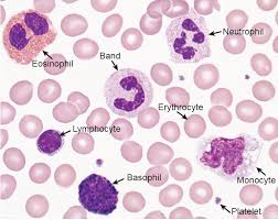
Morphology of Normal Blood Cells
-
Red Blood Cells (RBCs) – Erythrocytes:
-
Shape: Biconcave disc shape.
-
Size: 7-8 µm diameter.
-
Color: Pink with central pale area due to absence of nucleus. Contains hemoglobin.
-
Function: Oxygen aur carbon dioxide ka transport.
-
-
White Blood Cells (WBCs) – Leukocytes:
-
Neutrophils:
-
Size: 12-15 µm.
-
Nucleus: Multi-lobed (2-5 lobes).
-
Cytoplasm: Pale blue, fine granules.
-
Function: Phagocytosis aur bacterial defense.
-
-
Lymphocytes:
-
Size: 7-9 µm.
-
Nucleus: Large, round, occupying most of the cell.
-
Cytoplasm: Scanty, pale blue.
-
Function: Immunity aur antibody production.
-
-
Monocytes:
-
Size: 12-17 µm.
-
Nucleus: Large, kidney-shaped.
-
Cytoplasm: Abundant, pale blue.
-
Function: Phagocytosis aur antigen presentation.
-
-
Eosinophils:
-
Size: 12-17 µm.
-
Nucleus: Bi-lobed.
-
Cytoplasm: Bright orange-pink granules.
-
Function: Defense against parasites aur allergic responses.
-
-
Basophils:
-
Size: 12-17 µm.
-
Nucleus: Bi-lobed ya S-shaped.
-
Cytoplasm: Large, purple granules.
-
Function: Histamine release in allergic reactions.
-
-
-
Platelets – Thrombocytes:
-
Size: 2-4 µm.
-
Shape: Irregular, disc-like fragments.
-
Cytoplasm: Blue with purple granules.
-
Function: Blood clotting aur hemostasis.
-