Introduction
- Chromosomes ek thread-like structure hote hain jo har living cell ke nucleus me paaye jaate hain. Ye mainly DNA aur proteins (histones) se bane hote hain. DNA me genes hote hain jo humare traits (jaise eye color, height, blood group) aur body ke functions control karte hain.
- Human cells me normally 46 chromosomes (23 pairs) hote hain. Inme se 22 pairs autosomes hote hain aur 1 pair sex chromosomes hote hain (female = XX, male = XY).
- Cell division ke time, chromosomes ensure karte hain ki genetic material sahi tareeke se parent cell se daughter cells me transfer ho.
- Is tarah chromosomes genetic information ko store, protect aur next generation tak transmit karte hain.
Structure of Chromosomes
Chromosomes ekdum condensed thread-like structures hote hain jo DNA aur proteins se bane hote hain, aur nucleus ke andar genetic information carry karte hain.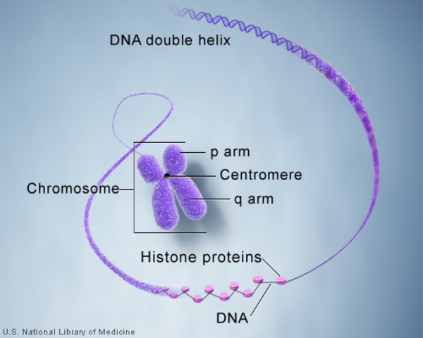
-
Composition:
-
DNA (~40%) → Genetic code rakhta hai.
-
Histone proteins (~50%) → Basic proteins (H1, H2A, H2B, H3, H4) jo DNA ko nucleosomes me package karte hain.
-
Non-histone proteins (~10%) → Enzymes, regulatory proteins.
-
RNA → thoda amount regulatory kaam ke liye.
-
-
Levels of packaging:
-
DNA double helix → 2 nm.
-
Nucleosome → DNA wrapped around histone octamer (“beads on string”).
-
30 nm fiber → aur zyada coiling.
-
Chromatin loops → 300 nm.
-
Fully condensed metaphase chromosome → 1400 nm.
-
-
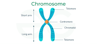
Parts:
-
Chromatid → Ek duplicated chromosome ki do identical halves.
-
Centromere → Constriction region jo spindle fibers se attach hota hai.
-
Telomere → DNA ke repetitive sequences (TTAGGG) jo chromosome ko protect karte hain.
-
Secondary constriction / NOR → rRNA genes ka region (nucleolus banata hai).
-
Satellite bodies → Chhoti chromatin masses jo secondary constriction se judti hain.
-
-
Types (centromere position ke basis pe):
-
Metacentric – centromere beech me.
-
Submetacentric – thoda side me.
-
Acrocentric – ek end ke paas.
-
Telocentric – bilkul end me (humans me nahi, rodents me).
-
Number of Chromosomes
-
Har species ka apna fixed chromosome number hota hai.
-
Examples:
-
Human → 46 (23 pairs).
-
Dog → 78.
-
Drosophila (fruit fly) → 8.
-
Onion → 16.
-
-
Diploid (2n) → Pura set (somatic cells).
-
Haploid (n) → Half set (gametes).
-
Abnormalities:
-
Aneuploidy → ek ya do chromosome extra/less (Down syndrome = Trisomy 21).
-
Polyploidy → complete set extra (plants me common).
-
Sex Chromosomes
-
Humans me 22 pairs autosomes + 1 pair sex chromosomes hote hain.
-
XX = Female, XY = Male.
-
Y chromosome me SRY gene hota hai jo male development start karta hai.
-
Disorders:
-
Turner syndrome (45,X).
-
Klinefelter syndrome (47,XXY).
-
Triple X syndrome (47,XXX).
-
XYY males (47,XYY).
-
Human Karyotype
-
Karyotype = poora chromosome set arranged in size order.
-
Female → 46,XX.
-
Male → 46,XY.
-
Preparation steps:
-
Dividing cells collect karo.
-
Colchicine se metaphase me arrest karo.
-
Hypotonic solution se swell karvao.
-
Fixation + staining.
-
-
Use: genetic disorders ka diagnosis (Down, Patau, Edwards), prenatal testing, cancer cytogenetics.
Methods for Chromosome Analysis
-
Classical: Karyotyping, banding.
-
Modern molecular: FISH, CGH, aCGH, microarray.
-
Flow cytometry: DNA content aur ploidy check karne ke liye.
-
NGS: genome-wide study.
Chromosome Banding
-
Chromosomes ko identify karne ke liye special stains.
-
Types:
-
G-banding → Giemsa stain, AT-rich dark bands.
-
Q-banding → Quinacrine fluorescence.
-
R-banding → reverse of G-banding.
-
C-banding → centromere region.
-
T-banding → telomere region.
-
-
Importance: structural abnormalities (deletion, duplication, translocation) detect karna.
FISH
-
Principle: Fluorescent DNA probes apne complementary sequence pe bind karte hain chromosome pe.
-
Types of probes:
-
Locus-specific.
-
Centromere-specific.
-
Whole chromosome painting.
-
-
Uses:
-
Microdeletions (DiGeorge syndrome).
-
Oncogene amplification (HER2 breast cancer).
-
Translocations detection.
-
Rapid prenatal diagnosis.
-
Comparative Genomic Hybridization
-
Principle: Test DNA aur reference DNA alag fluorescent dyes se label karke hybridize karte hain normal chromosomes/microarray pe.
-
Use:
-
Copy number variations detect karna.
-
Tumor DNA changes identify karna.
-
-
Limitation: balanced rearrangements (translocation, inversion) detect nahi hote.
Flow Cytometry
-
Principle: DNA-binding dye se cells stain karo → laser se pass karte hi DNA content measure hota hai.
-
Applications:
-
Cell cycle study (G1, S, G2/M phases).
-
Ploidy (diploid/aneuploid).
-
Oncology me tumor analysis.
-
Immunophenotyping.
-
Cell Cycle
-
Phases:
-
G1 → Growth, RNA/protein synthesis.
-
S → DNA replication.
-
G2 → Preparation for mitosis.
-
M → Mitosis + cytokinesis.
-
G0 → Resting state.
-
-
Regulation: cyclins + CDKs, checkpoints (G1/S, G2/M, spindle).
-
Error = cancer.
Mitosis
-
Purpose: Growth, repair.
-
Result: 2 diploid cells (identical).
-
Stages:
-
Prophase → Chromosome condense, spindle form.
-
Metaphase → Alignment at equator.
-
Anaphase → Sister chromatids separate.
-
Telophase → Nuclear envelope reappear.
-
Cytokinesis → Cytoplasm division.
-
-
Error → Aneuploidy, tumor.
Meiosis
-
Purpose: Gamete formation.
-
Result: 4 haploid cells (genetically different).
-
Meiosis I (Reductional division):
-
Homologous chromosome pairing (synapsis).
-
Crossing-over (genetic recombination).
-
Homologs separate → 2 haploid cells.
-
-
Meiosis II (Equational division):
-
Sister chromatids separate → 4 haploid gametes.
-
-
Significance:
-
Genetic diversity.
-
Chromosome number maintain.
-
Error → Nondisjunction (Down, Turner, Klinefelter).
-