Principle
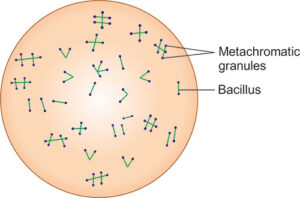
- Albert’s stain ka principle is based on Corynebacterium species ke granules ko stain karne ki ability.
- Yeh granules polyphosphate ke accumulation ka result hote hain.
- Albert’s staining method special dyes ka use karti hai jo in granules ko bind karte hain, unhe blue-black rang de dete hain, jabki puri bacterial cell ko greenish hue milti hai.
- Albert’s staining mein ek combination of dyes use hoti hai jo bacterial cells ko aur unke granules ko selectively stain karti hai.
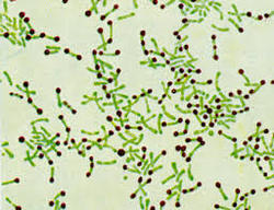
- Corynebacterium species ke metachromatic granules microscope par blue-black dikhte hain, jo unhe baaki bacteria se easily distinguish karne mein madad karte hain.
Requirements
Albert’s staining ko perform karne ke liye aapko yeh materials chahiye honge:
-
Microscope: Ek compound light microscope, preferably oil immersion lens (100x), taaki aap granules ko clearly dekh sakein.
-
Glass slides: Jisme bacterial smear prepare kiya jaa sake.
-
Inoculating loop: Bacterial sample ko slide par transfer karne ke liye.
-
Heat source: Smear ko slide par fix karne ke liye.
-
Staining rack: Slides ko staining ke dauran rakhne ke liye.
-
Distilled water: Slides ko rinse karne ke liye.
-
Staining containers: Staining reagents ko store karne ke liye.
Reagents
-
Albert’s Stain Solution: Yeh ek mixture hota hai do main components ka:
-
Alberts’ Solution A: Isme methyl violet aur iodine hoti hai, jo bacteria ko stain karti hai aur granules ko bind karti hai.
-
Alberts’ Solution B: Isme potassium iodide hota hai, jo iodine staining mein madad karta hai aur granules ka color enhance karta hai.
-
-
Acid-alcohol solution: Yeh decolorizing agent hota hai, jo non-granular areas se excess stain ko remove karta hai.
Sample
-
Bacterial Culture: Albert’s stain ka use zyada tar Corynebacterium diphtheriae ko detect karne ke liye kiya jaata hai. Sample kuch bhi ho sakta hai, jaise:
-
Throat swabs: Diphtheria ke suspected cases ke liye.
-
Tissue biopsies: Corynebacterium species ke infections ko diagnose karne ke liye.
-
Sputum aur other respiratory samples: Jahan patient ko respiratory symptoms ho jo diphtheria ke ho sakte hain.
-
-
Smear Preparation: Bacteria ka thoda sa sample slide par rakha jaata hai, aur usko ek thin, even layer mein spread kiya jaata hai. Phir smear ko air-dry karne ke baad heat-fix kar liya jaata hai taaki bacteria slide par chipak jaayein.
Procedure
-
Prepare the smear:
-
Bacterial sample ka ek chhota drop clean glass slide par rakhein.
-
Inoculating loop ka use karke sample ko evenly spread kar lein.
-
Smear ko air-dry hone dein aur phir slide ko flame ke upar 2-3 baar pass karke heat-fix kar lein.
-
-
Stain with Albert’s Solution A:
-
Albert’s Solution A (methyl violet aur iodine mixture) ko smear par kuch drops dal kar smear ko completely cover kar lein.
-
Stain ko 5-10 minutes ke liye room temperature par chhod dein.
-
-
Decolorize with acid-alcohol:
-
Acid-alcohol solution se slide ko gently rinse karein, excess stain ko remove karte hue, lekin granules ko intact rakhte hue.
-
Distilled water se rinse kar ke decolorization process ko stop kar dein.
-
-
Apply Albert’s Solution B:
-
Albert’s Solution B (potassium iodide solution) ko smear par apply kar ke 5 minutes tak chhod dein.
-
Yeh step granules ko zyada intense color deta hai aur unki visibility improve karta hai.
-
-
Final rinse and drying:
-
Distilled water se slide ko rinse karein aur extra stain ko remove kar dein.
-
Slide ko air-dry hone dein.
-
-
Microscopic examination:
-
Slide ko compound light microscope se examine karein, oil immersion lens ka use karte hue.
-
Granules dark blue-black dikhai denge, jabki bacterial cell ka baaki hissa greenish dikhai dega.
-
Results
-
Positive Results:
-
Bacteria mein characteristic metachromatic granules dark blue-black dikhai denge.
-
Bacterial cell ka baaki hissa greenish hoga.
-
Corynebacterium diphtheriae aur dusre Corynebacterium species ke granules easily visible honge.
-
-
Negative Results:
-
Agar bacteria mein metachromatic granules nahi hain, toh granules ka dark staining nahi hoga.
-
Cells greenish dikhai denge, bina kisi dark inclusions ke.
-
Applications
-
Diphtheria Diagnosis: Albert’s staining ka primary use Corynebacterium diphtheriae ko identify karne ke liye hota hai. Metachromatic granules ka presence diphtheria ko confirm karne mein madad karta hai.
-
Identification of Corynebacterium Species: Albert’s staining dusre Corynebacterium species ko bhi identify karne mein madad karta hai, jo opportunistic infections cause karte hain.
-
Bacterial Research: Yeh method Corynebacterium species ka study karne aur unke morphology aur granule formation ko samajhne mein use hota hai.
-
Educational Purpose: Albert’s stain microbiology labs mein students ko bacterial granules dikhane ke liye use hota hai, jo unke understanding ko enhance karta hai.
-
Confirmation of Infection in Clinical Specimens: Yeh effective method hai diphtheria aur dusre Corynebacterium infections ko confirm karne ke liye throat swabs aur clinical samples mein.