Introduction
-
Histopathology = Tissues ka microscope ke through study, disease ko detect karne ke liye.
-
Cytopathology = Cells ka study microscope ke under.
Ye dono disease detection, cancer diagnosis, prognosis aur research ke liye bahut important hain.
Lab me examination ke do main tareeke hote hain:
-
Tissue Examination (Histopathology)
-
Cell Examination (Cytopathology)
Tissue Examination
Yahaan pura tissue sample ko process karke examine kiya jata hai.
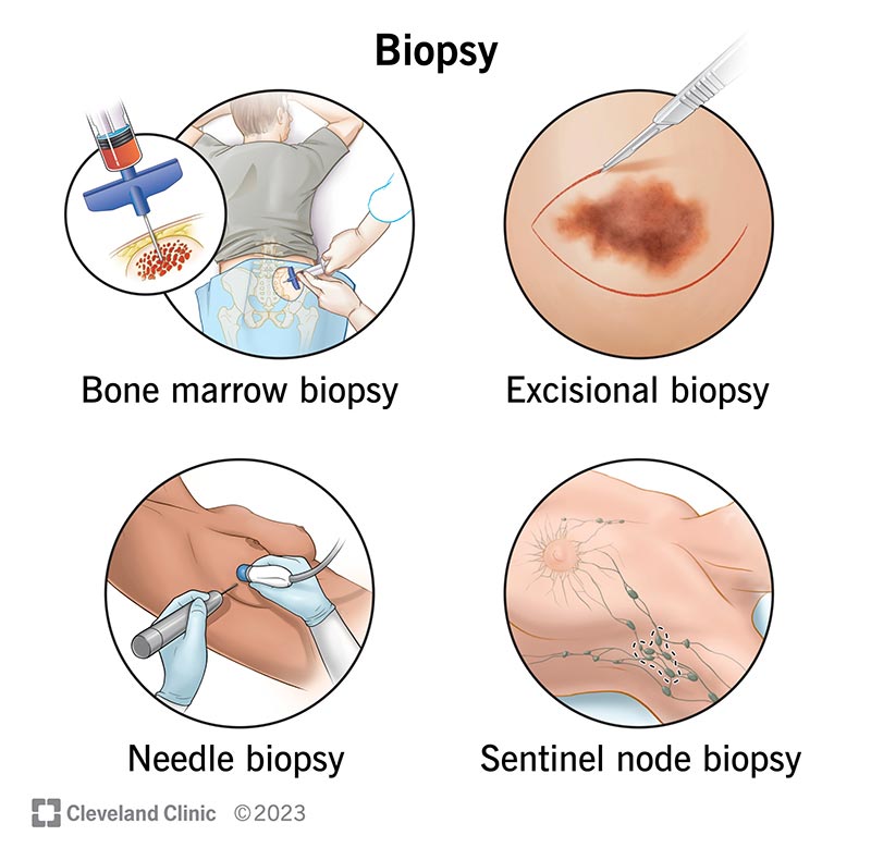
A. Biopsy
-
Matlab: Zinda patient se chhota tissue piece nikalna diagnosis ke liye.
-
Types:
-
Incisional biopsy – sirf lesion ka ek hissa liya jata hai.
-
Excisional biopsy – pura lesion nikal diya jata hai.
-
Needle biopsy – needle se chhota tissue sample liya jata hai.
-
Endoscopic biopsy – endoscope se (jaise stomach, colon).
-
-
Use: Cancer, infection, chronic disease ka diagnosis.
B. Surgical Specimen
-
Surgery ke time nikale huye organs/tissues.
-
Example: Appendix, Gall bladder, Breast lump.
-
Pehle gross examination hota hai (size, shape, weight, margins).
-
Fir microscopic exam ke liye process kiya jata hai.
C. Autopsy
-
Dead body se tissue ka examination.
-
Types:
-
Clinical autopsy (cause of death ke liye).
-
Medico-legal autopsy (suspicious cases).
-
-
Disease ke natural history aur treatment response samajhne me help karta hai.
D. Tissue Processing
Tissue ko microscope ke liye tayaar karna:
-
Fixation → Formalin me dal kar tissue ko preserve karte hain.
-
Dehydration → Alcohol series me dal kar paani nikalte hain.
-
Clearing → Xylene se alcohol replace karte hain.
-
Embedding → Paraffin wax block banate hain.
-
Sectioning → Microtome se 3–5 micron thin sections banate hain.
-
Staining → Hematoxylin & Eosin (H&E) sabse common.
E. Special Stains
-
PAS → glycogen, mucin.
-
Ziehl–Neelsen → TB bacilli.
-
Silver stain → fungus, reticulin fibers.
-
Congo red → amyloid (apple-green birefringence).
-
Masson’s Trichrome → collagen.
F. Immunohistochemistry
-
Antibody–antigen reaction ke zariye proteins ko detect karna.
-
Example:
-
Breast cancer me ER, PR, HER2.
-
Ki-67 proliferation marker.
-
-
Diagnosis + prognosis + treatment ke liye useful.
G. Frozen Section
-
Tissue ko cryostat me turant freeze karke section banate hain.
-
Surgery ke time quick diagnosis ke liye use hota hai.
-
Example: Tumor margin check karna.
H. Electron Microscopy
-
Bohot high magnification pe organelles, viruses, basement membrane details dikhata hai.
-
Renal biopsy, muscle diseases, viral studies me kaam aata hai.
Cell Examination
Yahaan sirf cells ka study hota hai, tissue architecture preserve nahi hota.
A. Exfoliative Cytology
-
Jo cells naturally shed hote hain unka study.
-
Example: Pap smear – cervical cancer screening.
-
Aur examples: Sputum cytology, Urine cytology.
B. Fine Needle Aspiration Cytology (FNAC)
-
Fine needle se lump ya swelling se cells nikal kar slide par smear banate hain.
-
Stains: Papanicolaou, Giemsa.
-
Advantages: Quick, cheap, OPD me possible.
-
Use: Breast, thyroid, lymph node, salivary gland lumps.
C. Body Fluids Cytology
-
Fluids jaise Pleural, Peritoneal, CSF, Synovial fluid ka exam.
-
Malignant cells ya infections detect karte hain.
D. Imprint Cytology
-
Fresh tissue ko slide par press karke imprint banate hain.
-
Rapid diagnosis in lymph node, breast, thyroid.
E. Cytochemistry
-
Special stains ka use chemical nature dekhne ke liye.
-
Example:
-
PAS → glycogen.
-
Sudan Black → lipids.
-
Peroxidase stain → myeloid cells.
-
F. Flow Cytometry
-
Cell suspension ko laser ke through pass karte hain.
-
Antibody markers detect karne ke liye fluorescent tags use hote hain.
-
Uses: Leukemia/lymphoma classification, minimal residual disease detection.
Tissue vs. Cell Examination: Comparison
| Feature | Tissue (Histopathology) | Cell (Cytopathology) |
|---|---|---|
| Sample | Biopsy, surgical specimen | FNAC, Pap smear, body fluid |
| Architecture visible? | Yes (tissue structure intact) | No (single cells only) |
| Time required | Long (processing chahiye) | Short (rapid results) |
| Common stains | H&E, special stains, IHC | Pap, Giemsa, cytochemistry |
| Major use | Tumor typing, staging, prognosis | Screening, quick diagnosis |
| Example | Breast carcinoma biopsy | FNAC of breast lump |